General Inspiration:
Bill Bryson – “The Body: A Guide for Occupants”
- All functions of the body and brain
- How he was interested in the scientists discovered and the stories of how the scientists discovered things, who they are, their circumstances when discovering the things
Microscopy – specific Inspiration “The Microscopists” by Howard Hughes Medical Institute
- Eric Betzig
- Jennifer Lippincott-Schwartz (takes canoe and crosses Atomic(??))
Yale West Campus
- Acquired by Yale in 2007
- 1.6 million square feet of lab space, conference rooms and dining area
- 1600 faculty, staff and students
- Seven institutes:
- Energy sciences
- Systems biology
- Bimolecular design and discovery (Chemical biology)
- Cancer biology
- Nanobiology
- Microbial sciences
- Preservation of cultural heritage
- From different fields, work together
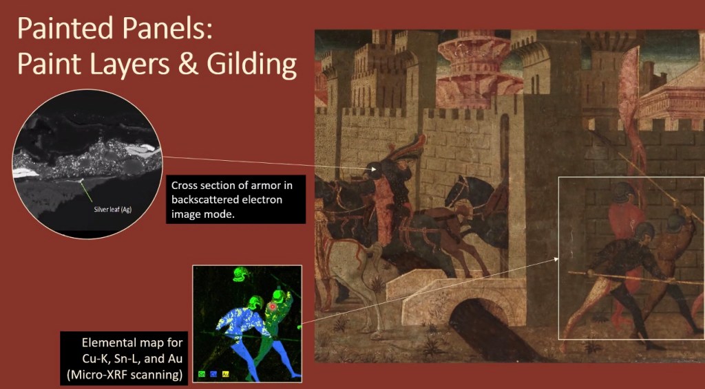
Cassone by Paolo Uccello
- Marriage chests, for rich couples because hand painted by famous artists
- Interesting for imaging because scientists work with conservators because the painting or the structure of the pieces of the art
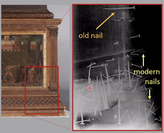
How do they preserve the old pieces of art while scanning? (I asked this question)
- They add a pink layer to fill in some empty spaces to help preserve the pieces
Core Facilities
Benefits:
- Can get state of the art facilities —> save money and continuously buy the latest equipment that there is (better microscopes and stronger technology)
- Great core technology educates next generation of scientists
- Technology industry have a strong interest in selling equipment and having their brand represented in different core facilities
- Can link cores together to work on uncertain projects
Imaging Core
- Every minute shake can give blurry image, films are on optical tables
- Basement is close to rock bottom of the structure —> fewer vibrations
- Avoid interference of sunlight
Sol LeWitt – gave descriptions to his art pieces (didn’t paint them himself)

Sample —> visualize it first —> have image on microscope —> describe it through qualitatively/quantitatively
- Stereomicroscope (prep samples to go to real microscope, check the microscope objectives, doesn’t need to be on optic table)
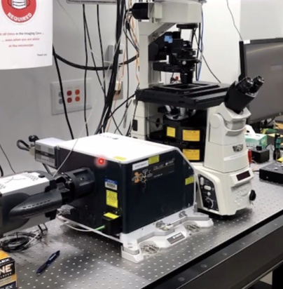
- Widefield Fluorescence Microscope – floating on optical table
- Cell culture fluorescent
How does fluorescence work?
= Jablonski diagram shows electron in ground and excited
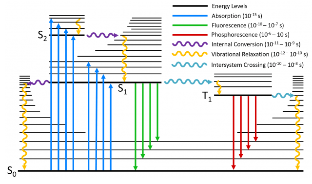
Before emission light hits, the electrons are in ground state. The light carries some energy, which pushes the electrons from ground to excited state. As the electron drops down to the bottom level of the excited state, it emits light (which is a different wavelength since each visible spectrum is a different color), lose energy to emit color, and returns to ground state.
Laser Microdissection Microscope
- Need a very thin samplle (couple of microns thick)
- Put on slide and on the stage of microscope
- Attached skin monitor, once you visualize, you can draw a certain shape
- Certain laser on microscope that allows you to cut the way you want
- Precision depends on laser and strength of laser (stronger laser —> less precision)
- Cutting salad with knife vs chainsaw
- Attached skin monitor, once you visualize, you can draw a certain shape
Using this microscope, can print proteins from protein database, want to visualize how proteins interact with each other, can see
Spinning disk confocal
- Microscope disk spins really fast to produce image with laser
Optical Tweezers

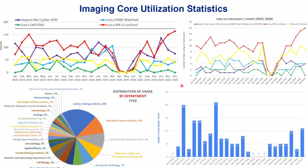
Imaging core utilization statistics
Projects in the Imaging Core
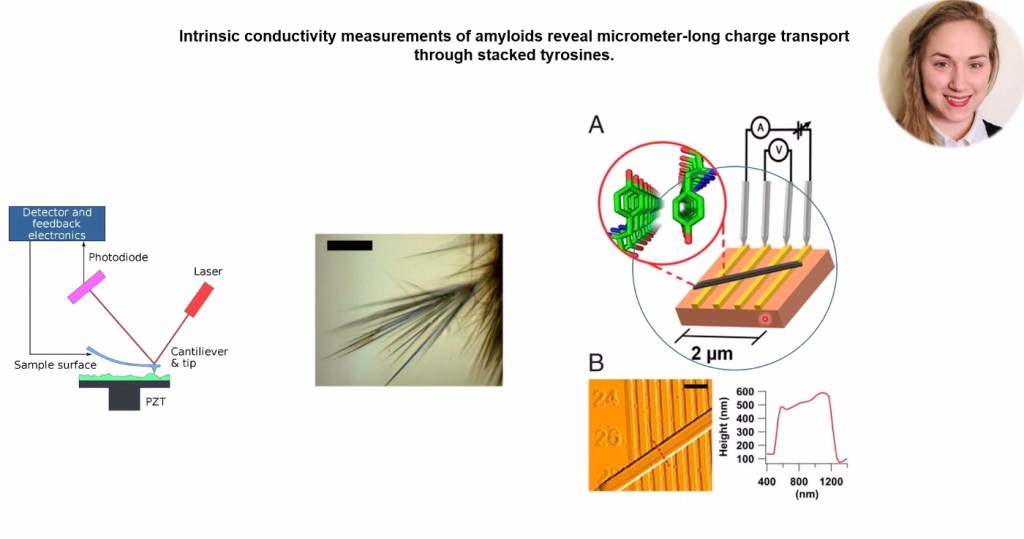
- Bacteria and how they transport charges
- Needles made of specks of amino acids that can transport charges
Needles placed on electrodes.
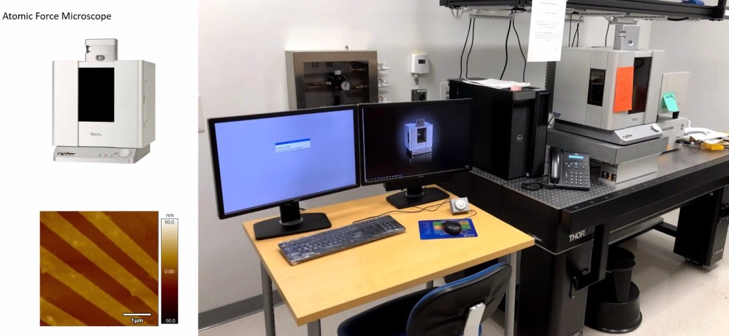
Process: AFM works by dragging tip across high profile of sample surface (measuring the changes in the height by pointing a laser at the tip and look at reflection on photodiode) and when tip moves —>> heigh changes —> reflection varies -> computer able to predict precise high profile
Another technology:
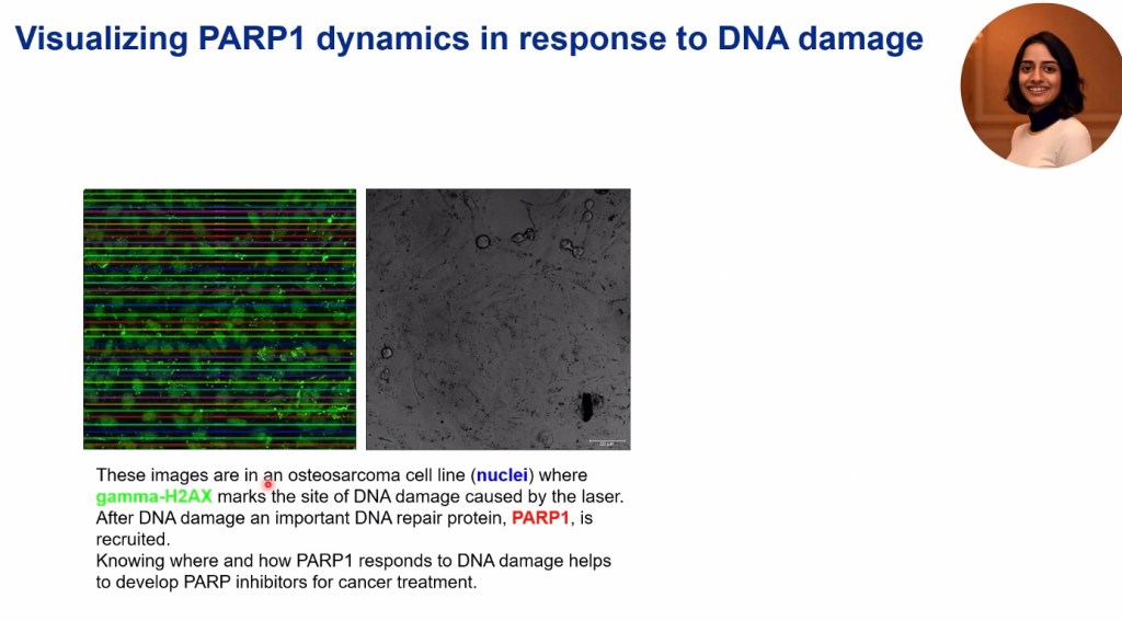
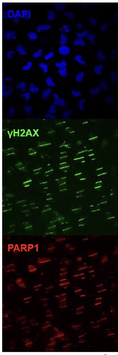
make damage and go after a minute and image it, see how fast the proteins get recruited —> to cure cancer that damage will produce
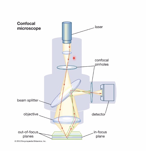
Excite the four floors, follow red area, go through objective through another pin hole (in focus so goes through pin hole through detector)
Out of focus does go through direction of pinhole but not so precisely (only in focus pinhole)
Turning intensity up can produce different images
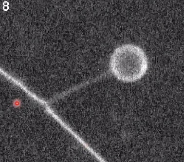
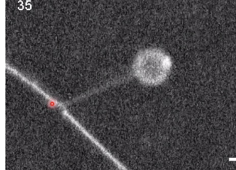
Optical tweezers
= Cell membrane sticks to it, briefly remove the bead —> creates a membrane tether (high urbiture of tether —> use it to see curvature preference of proteins)
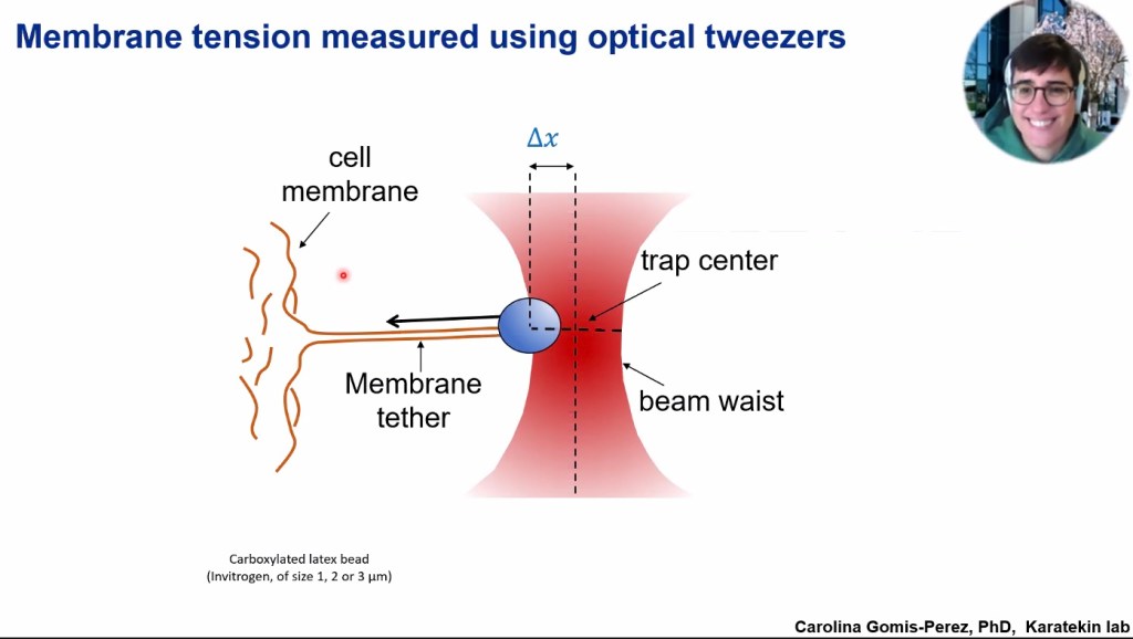
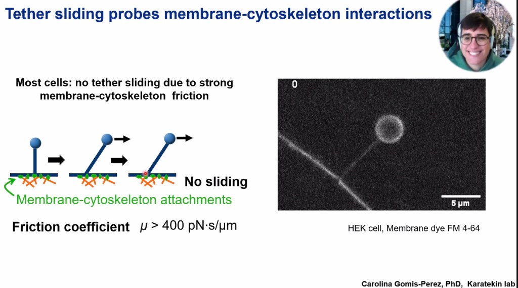
Underneath the membrane are the orange bits (membrane cytoskeleton)
Pull tether upward, pull bead to side. Will base of tether move with the beat when its dragged in one direction or stay put?
Exocytosis (cell that has a molecule stored in a vessicle inside the cell) and to fire the synapse, bring vesicle to membrane, opens the pore and releases neurotransmitter into atmosphere
- Two membrane need to fuse together to create a not leaking vesicle outside the cell
- Does the membrane tension in this cell memebrae affect how the release
- Pore is formed to rip this open and things are released but if there’s high tension, hard to perform the pore as they’re single lipids
- Does the membrane tension in this cell memebrae affect how the release
Goldfish retinal bipolar neuron —> want to study in synapses that will transmit signals from one neural cell to another (usually chemical synapses are super tiny, smaller than the cell, usually 200 nanometers), hard to visualize them
Terminal is 10 nano meters (much smaller than usual cells)
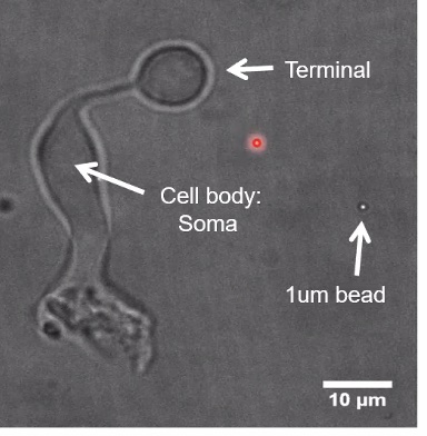
Base of tether actually moved
(None of the HeLA cells slide but 80% of terminal tether slide)

Advanced three-dimensional imaging of embryos
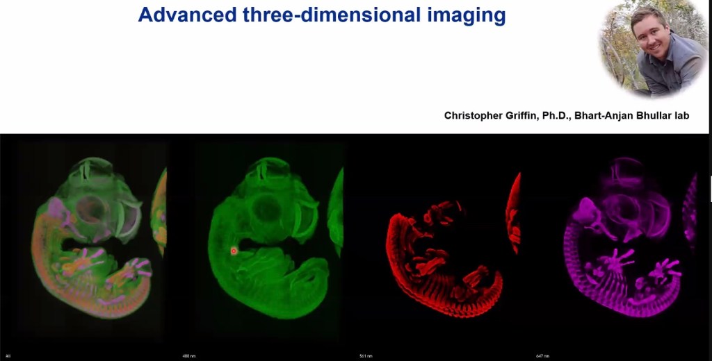
Embryo of a vertebrate (parrot), look at different cellular structures
- Takes several weeks to do procedure
- Makes sample transparent
- Embryo is a centimeter in size —> light wouldn’t get far, would be reflected
- To scan whole embryo, need to make ti transparent
- Because scan with high magnification, tile scan that scans each individual slide smaller than whole image (image whole image in 2d and different layers of embryo for 3d image)
- Makes sample transparent
– Joanna Kim, August 1rst, 6:07PM KST –
